Description
HardyCHROM UTI, a chromogenic medium for all urinary tract pathogens and other uses, 15x100mm plate, order by the package of 10, by Hardy Diagnostics.
One plate does it all!
HardyCHROM™ UTI is a differential culture medium that facilitates the isolation and differentiation of urinary tract pathogens, including gram-negative and gram-positive bacteria. The development of various colors, due to chromogenic substances in the media, allows for the differentiation of microorganisms from the primary set-up of a urine specimen.
There are a number of organisms routinely isolated from urinary tract infections (UTI). Most UTIs are caused by Escherichia coli alone, or in combination with other organisms. The most frequently isolated species produce characteristic enzymes. Chromogenic substrates (chromogens) incorporated into HardyCHROM™ UTI produce different colored compounds when they are degraded by specific microbial enzymes. Thus, HardyCHROM™ UTI can be used for the cultivation and differentiation of various groups of organisms with only a minimum number of confirmatory tests.
This media is to be stored and incubated in the dark. If you have a light in your refrigerator, incubator, or you leave the media exposed to light on the bench top, Hardy Diagnostics offers free Blok-Boxes. Blok-Box is a reusable cardboard box that holds one sleeve of ten petri plates.
Features and Benefits:
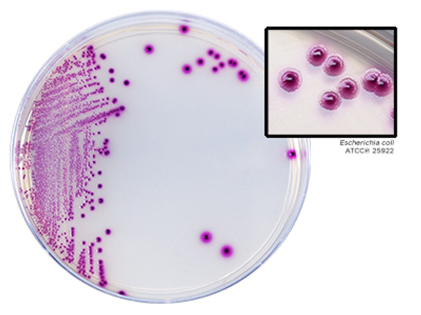
- E. coli produces large magenta colonies (confirmatory, no further testing required)
- Enterococcus faecalis/faecium produces small, turquoise-colored colonies
- Pseudomonas spp. produce light yellow/green, translucent colonies.
- Klebsiella, Enterobacter, and Serratia spp. produce large, deep blue colonies
- Staphylococcus saprophyticus produces opaque, pink colonies
- Candida spp. produces small, white, moist colonies
- Proteus, Morganella, and Providencia spp. produce clear to light yellow colonies with a diffuse golden-orange halo in the medium
- Staphylococcus aureus produces opaque, white-colored colonies
- Citrobacter spp. produce dark blue colonies often with a rose halo in the surrounding media
INTENDED USE:
HardyCHROM UTI is a non-selective chromogenic medium recommended for the cultivation, differentiation and enumeration of urinary tract pathogens based on colony color and morphology.
SUMMARY:
There are a number of organisms routinely isolated from urinary tract infections (UTI). Most UTIs are caused by Escherichia coli alone, or in combination with other organisms. The most frequently isolated species produce characteristic enzymes. Chromogenic substrates (chromogens) incorporated into HardyCHROM™ UTI produce different colored compounds when they are degraded by specific microbial enzymes. Thus, HardyCHROM™ UTI can be used for the cultivation and differentiation of various groups of organisms with only a minimum number of confirmatory tests. Peptones supply the necessary nutrients, and the mixture of chromogens permits detection and differentiation of the isolated organisms. This medium contains no inhibitory substances and is not selective. The swarming of Proteus is partially to completely inhibited.
FORMULA:
Ingredients per liter of deionized water:* Peptones 16.0gm Chromogenic Mixture 5.0gm Agar 15.0gm Final pH 6.9 +/- 0.3 at 25°C. * Adjusted and/or supplemented as required to meet performance criteria.
STORAGE AND SHELF LIFE:
Storage: Upon receipt store at 2-8°C away from direct light. Media should not be used if there are any signs of deterioration (shrinking, cracking, or discoloration), contamination, or if the expiration date has passed. Chromogens are especially light and temperature sensitive; protect from light, excessive heat, moisture, and freezing. The expiration date applies to the product in its intact packaging when stored as directed. This product has the following shelf life from the date of manufacture: 90 Days: G313 HardyCHROM™ UTI
PRECAUTIONS:
This product may contain components of animal origin. Certified knowledge of the origin and/or sanitary state of the animals does not guarantee the absence of transmissible pathogenic agents. Therefore, it is recommended that these products be treated as potentially infectious and handled observing the usual universal blood precautions. Do not ingest, inhale, or allow to come into contact with skin. 080715kdp HardyCHROM™ UTI Page 2 of 8 This product is for in vitro diagnostic use only and is to be used only by adequately trained and qualified laboratory personnel. Observe approved biohazard precautions and aseptic techniques. All laboratory specimens should be considered infectious and handled according to "standard precautions".
PROCEDURE:
Specimen Collection: Consult listed references for information on specimen collection.(2-4) Infectious material should be submitted directly to the laboratory without delay and protected from excessive heat and cold. If there is to be a delay in processing, the specimen should be refrigerated until inoculation. Consult the listed references for information regarding the processing of specimens.(1-5) Protect media from light during storage and incubation as the product is light sensitive. Method of Use: Allow the plates to warm to room temperature. The agar surface should be to dry prior to inoculating. Inoculate and streak the specimen as soon as possible after collection. For quantitative testing streak plate with 0.01ml (Cat. no. HS10R) or 0.001ml calibrated loop (Cat. no. HS1R). Incubate plates in an inverted position, aerobically at 35 +/- 2°C for no less than 24 hours. Examine plates for colonies showing typical morphology and color after 24 hours, but no later than 36 hours. Do not incubate in an atmosphere supplemented with CO2.
INTERPRETATION OF RESULTS:
After incubation, the plates should show isolated colonies. Isolated colonies are necessary for demonstration of typical color and morphology. Escherichia coli produces medium to large sized colonies that are rose to magenta in color, with darker pink centers. No further testing is needed. Colonies that resemble E. coli (pink to rose), but are small or pinpoint in size, require further identification procedures such as the Indole Spot Test (DMACA, Cat. no. Z65). See "Limitations" section below. Enterococcus spp. appear as small, teal to turquoise colored colonies. No further testing is needed. Proteus, Morganella, and Providencia spp. produce clear to light yellow colonies with golden-orange halo diffused through surrounding media. Additionally, approximately 50% of Proteus vulgaris isolates will produce blue-green or green colonies with a golden-orange halo. Proteus vulgaris can be identified by a positive spot indole test (Cat. no. Z65). Further biochemical tests are needed for complete identification of the other members of this group. Indole Spot Test (Cat. no. Z65) may be performed from the plate. H2S production and ornithine decarboxylase (Cat. no. Y44) permit differentiation of the genera. Klebsiella, Enterobacter, and Serratia spp. produce large, deep blue or dark indigo colonies. Citrobacter spp. produce dark blue colonies often with a rose halo in the surrounding media. Further biochemical tests such as the Microgen GNA Panel (Cat. no. MID64) are needed for complete identification. Pseudomonas spp. produce colorless to light yellow-green, translucent colonies which may have a slight iridescence with crinkled edges. Further biochemical tests, including an oxidase test (Cat. no. Z93) may be needed for complete identification. 080715kdp HardyCHROM™ UTI Page 3 of 8 Staphylococcus aureus produce opaque, cream to white colored colonies. Note: Colonies may turn pink after 72 hours. Further tests (StaphTEX™, Cat. no. ST50) are needed for complete identification. Staphylococcus saprophyticus produce opaque, pink colonies. Further tests, such as novobiocin-resistance (Cat. no. Z7291), are needed for complete identification. Staphylococcus epidermidis grows as small, white colonies. Further biochemical tests are needed for complete identification. Candida albicans, Candida tropicalis, and Candida glabrata produce small, opaque, white, moist colonies. Further biochemical tests such as AlbiQuick™ (Cat. no. Z121) or HardyCHROM™ Candida (Cat. no. G301) are needed for complete identification. Candida krusei appears as small, white, dry colonies which have a rough appearance. Further biochemical tests are needed for complete identification. Listeria monocytogenes or other Listeria spp. may be present in urine. Colonies of Listeria are very small, blue to bluegreen colonies. Perform a Gram stain of organisms producing small, blue to blue-green colonies that are PYR-negative. The presence of gram-positive bacilli is suggestive of Listeria spp. but further biochemical tests are necessary for complete identification. Streptococcus agalactiae isolated from urine appears as very small clear blue colonies, very small clear white colonies or very small pink or pink-blue colonies. Further tests, such as Strep B Carrot Broth™ (Cat. no. Z140), are needed for complete identification. For organisms other than E. coli and Enterococcus spp. biochemical tests should be performed on colonies from pure cultures for complete identification. Use a filter paper to perform rapid tests. Do not apply any detection reagents directly on the colonies growing on the medium.


